X-ray Microscopy
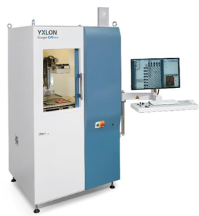
The Yvlon Cougar X-ray microscope provides non-destructive imaging capabilities on specimens across a range of length scales, observing features with sizes spanning from millimeters to micrometers.
The techniques reveal structural characteristics and flaws, such as cracks and pores, or composition features. There is a wide variety of applications in materials science, life science, geoscience and microelectronics - from biological objects such as insects or bones, over structural materials like alloys - to image the distribution of precipitates - and composites - to image the components and the quality of interfaces such as interconnects in micro-electronic products and miniaturized sensors.
Typical Applications:
- Inspect BGA and QFN attachment, solder shorts, PTH filling and detect counterfeit components
- Failure analysis of high-resolution bond wire and package level inspection
Specifications:
- X-ray tube: open tube 25kV to 160kV, 0.01mA to 1mA
- Maximum tube power: 64W
- Maximum target power: 10W
- Detail detectability: < 0.75 um
- Spatial resolution: 1.5um
- 5-axis (X, Y, Z tube (motorized), Z Detector (motorized), Tilt Detector (motorized)) manipulation
- geometric/ total magnification: 2,000x/ 10,000x
- CMOS Detector with 50mm x 50mm, 1,012x1,012 pixel, pixel size 50um, frame rate: max. 30 fps
- Oblique viewing by tilting detector +/- 70o
- focus-object distance (FOD): 0.25mm
- focus-detector distance (FDD): 50mm
- Inspection map stitching feature
- Void calculation feature.
- CT function to switch from 2D radioscopy to 3D uCT with real-time 3D volume rendering and 3D visualization
- 3D analysis
- Maximum inspection area: 310mm x 310mm
- Maximum sample weight: 5kg